(D) = True - Radiographs reveal Post-Axial Polydactyly, Sub-Type A and one should proceed with surgical resection of the most lateral digit.
Post-Axial Polydactyly is the most common polydactyly variant and correlates with a lateral digit duplication. The classification system developed by Temtamy and McKusick based on pedigree analysis, classified polydactyly of the hand into anatomic and genetic grounds (adapted from McGlamery's here for the foot categories consisted of Post-Axial and Pre-Axial.
(1) Post-Axial Polydactyly correlates with hyperdactyly that denotes a lateral digital duplication.
-
Sub-Type A = well-formed, articulated digit
-
Sub-Type B = rudimentary digit often without skeletal component
(2) Pre-Axial Polydactyly is applied to medial digit duplication.
(3) Central Polydactlyly is the least common and is a duplication of the innermost digit.
In regards to surgical intervention, the best procedure is one that will give the most normal shape and function to the forefoot. Given the appearance and function, it is recommended that the lateral-most digit an Post-Axial Polydactyly and the medial-most digit in Pre-Axial Polydactyly is excised
to allow for a straight anatomic contour of the forefoot.
In the image above the patient presents with a Post-Axial Polydactyly Sub-Type A, and the best surgical decision would be resection of the lateral most fifth digit.
Citation:
-
Filiatraut, A. D. (2013). Congenital Digital Deformities: Polydactyly. In McGlamry's comprehensive textbook of foot and ankle surgery (pp. 1097-1105). Atlanta, GA: Wolters Kluwer Health/Lippincott Williams & Wilkins.

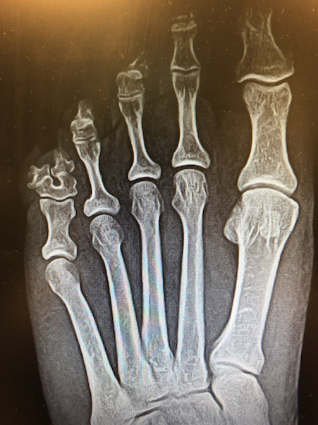 Photo credit by Michael E. Calderone, DPM